Showing posts with label Retinal. Show all posts

Friday, May 19, 2017

NeoVista Epi Retinal Strontium 90 Treatment for AMD Update 3


During the 2008 Retina Society Meeting, held last weekend, NeoVista, Inc. provided eighteen-month data from its Phase II feasability study of the company’s novel beta radiation epi-retinal therapy for the treatment of the wet form of age-related macular degeneration (AMD). The long-term data from the study, which was initiated to test the safety and efficacy of their therapy when used in conjunction with Avastin (bevacizumab), showed a marked advancement in mean visual acuity results at month 18, while only a limited number of patients required additional injections of Avastin.

The data were presented at the Retina Society Meeting by Nelson R. Sabates, MD, Professor and Chairman, Department of Ophthalmology, University of Missouri-Kansas City (UMKC) School of Medicine and the lead investigator in NeoVista's ongoing Phase III study, CABERNET (CNV Secondary to AMD Treated with BEta RadiatioN Epiretinal Therapy).

"The data released demonstrate that NeoVista's concomitant approach has the potential to offer patients a less frequent treatment option that is just as effective, if not more effective, than the current standard of care," said Dr. Sabates. "It's highly encouraging to continually see patient outcomes improving as the study progresses."

"We're very delighted with the latest data from our Phase II study, as not only did the visual acuity improve in our patients over the long-term, but very few patients received additional injections as well," said John N. Hendrick, President and CEO of NeoVista. "The ultimate pledge of this therapy continues to be demonstrated as the long-term data hold promise in minimizing the treatment burden both for patients and physicians, not to mention the overall financial burden for the healthcare system."

NeoVista's revolutionary therapy applies a targeted dose of beta radiation to the leaking blood vessels that affect central vision; concomitantly, two injections of an anti-vascular endothelial growth factor (anti-VEGF) agent are delivered to maximize the acute therapeutic response. Preliminary data show that NeoVista's targeted radiation therapy can be safe for both the patient and the physician, and may be able to restore the patient's vision. The current standard of care for wet AMD requires persistent injections of anti-VEGF drugs for an indefinite period.

The ongoing multicenter feasibility study enrolled 34 trial participants (with a mean age of 72 years) from June 2006 to April 2007 at two centers in Brazil and one in Mexico. These patients, with predominantly classic, minimally classic, or occult (with no classic) choroidal neovascularization (CNV), received a single 24 Gy treatment of NeoVista's epiretinal brachytherapy in combination with two intravitreal injections of Avastin, one dose prior to or at the time of radiation delivery and another one month later, depending on which arm of the trial the patient was enrolled in. Additional therapy was delivered based upon the investigator's evaluation of disease activity.

Analysis of 18-month follow-up on the first 25 trial participants to reach that milestone, as shown in Graph 1 below, shows a mean improvement in visual acuity of 10.7 letters using the Early Treatment Diabetic Retinopathy Study (ETDRS) test; 96 percent of patients lost 15 letters or fewer, 76 percent gained some letters, 44 percent gained 15 or more letters, and 8 percent gained 30 or more letters. Of particular interest, 68 percent of the patients in the study did not require additional injections of Avastin throughout the 18-month period and the average number of additional injections within this subset was only 2.4 injections by month 18.


Graph 1

The visual acuity data after 18 months compares favorably with the results reported after 12 months, as shown in Graph 2 below. (This graph compares the Epi-Rad treatment without Avastin (purple color), with Epi-Rad plus Avastin (blue color) and the Marina (green) and Anchor (red) studies, which both used Lucentis.)

Graph 2

For more information on the NeoVista Epi-Retinal treatment, and for further information on the Marina and Anchor Studies, see my three earlier reports on NeoVista, posted November 19, 2007; July 11, 2007; and February 14, 2007.

Most of the limited number of adverse events were related to the vitrectomy procedure (retinal tear, retinal detachment, subretinal hemorrhage, and vitreous hemorrhage), rather than the epiretinal brachytherapy. To date, no instances of radiation toxicity have been reported by the Doheny reading center.

In contrast to other forms of radiation therapy for wet AMD, NeoVista's approach delivers the peak dose of energy directly to the lesion without damaging the normal retinal vasculature. Utilizing strontium 90, the focused energy is delivered to a target area up to 3 mm in depth and up to 5.4 mm in diameter. Importantly for patients, the systemic exposure to radiation is minimal, as the effective dose to the entire body from NeoVista's epiretinal device is less than that from a typical chest x-ray.

With the continued promise of these Phase II trial results, NeoVista continues to enroll patients in the company's pivotal trial, CABERNET. CABERNET is a multicenter, randomized, controlled study that will enroll 450 subjects at 45 sites worldwide, evaluating the safety and efficacy of NeoVista's epiretinal brachytherapy delivered concomitantly with the FDA-approved anti-VEGF therapy Lucentis (ranibizumab) versus Lucentis alone.


Wednesday, April 19, 2017

NeoVista Epi retinal Strontium 90 Treatment for Wet AMD


In my attempt to keep readers of this web site apprised of potential new treatments for age-related macular degeneration, I would like to present early positive results for a brand new treatment for AMD. The new vice president of sales & marketing at NeoVista, Tony Moses, an old friend, sent me a copy of the presentation of the recently presented first results for using strontium 90 in combination with a VEGF drug to alleviate vision loss in wet AMD. With his permission, here are the facts to date.

On January 16, 2007, Dr. Andrew Schachat, Vice Chair for Clinical Affairs, Vitreoretinal Department of the Cole Eye Institute, presented the first data on NeoVista’s Epi-Rad 90 Ophthalmic System at the Hawaiian Eye meeting (RHEM). The procedure involves the epi-retinal application of strontium 90 radiation for the treatment of wet AMD. A review of some of the early findings from his presentation are reported below.

Radiation treatment for AMD

NeoVista, Inc., of Fremont, Calif, released data demonstrating a potential benefit of treating wet AMD with radiation using the company's Epi-Rad 90 Ophthalmic System. Clinical data was presented from 2 separate feasibility studies that utilized the NeoVista product. The first study, involving a total of 24 patients, comprised 2 different doses of radiation (15 Gy and 24 Gy) delivered to the retina. The second study, which involved 20 patients, utilized a concomitant approach of 24 Gy radiation plus Avastin, where an injection of the drug was administered at the time of radiation treatment and an additional injection administered 30 days later. This is the approach the company plans to follow when it begins its pivotal 450-patient CABERNET trial this year. The company will utilize Lucentis instead of Avastin in this trial.

The Procedure and Early Results

The procedure involves performing a partial vitrectomy under local anesthesia. The delivery of the radiation takes between 3 and 5 minutes, depending on the calibration certification for the device being used, while the total procedure takes about approximately 40 minutes. The focal radiation penetration is about 3 mm into the choroid. The company claims that the ionizing radiation has a toxic effect on local pro-inflammatory and fibroblast cell populations, permanently disabling the proliferating CNV Cells.

In the two feasibility studies, using radiation alone, the lower dosage test (15 Gy) was not impressive, showing a loss in visual acuity after 9 (-2.4 letters) and 12 months (-3.2 letters).The study utilizing a higher dosage of strontium 90 (24 Gy) had better results, showing an increase in visual acuity after 9 (+5.6 letters) and 12 months (+7.6 letters). This compared favorably with the MARINA study of Lucentis, which had an increase of +7.2 letters after 12 months.


However, the second study, which utilized a concomitant approach of 24 Gy radiation plus Avastin, where an injection of the drug was administered at the time of radiation treatment and an additional injection administered 30 days later, was even more impressive. After only 2 and 3 months followup, the 20 patients showed an increase in VA of +14.2 and +14.9 letters, beating the MARINA study results by a wide margin.


Eugene de Juan, Jr., MD, the Jean Kelly Stock Professor of Ophthalmology at the University of California San Francisco and the inventor of the NeoVista treatment approach, commented, "Although the follow-up period (3 months) is relatively short, the results observed from the concomitant trial are extremely encouraging. The percentage of patients who improved in visual acuity by greater than 3 lines was reported at 50%, which is far above the 34% that was reported in the Lucentis MARINA Study. Granted, the NeoVista sample size is much smaller than that garnered from MARINA, but the evidence does support a closer investigation of this concomitant approach."

"We continue to see vision stability, and in many cases, vision improvement after just one treatment," stated John Hendrick, President and CEO of NeoVista. "The potential impact of our technology will greatly benefit patients, physicians, and the overall health care system. We will soon begin our Pivotal Trial incorporating concomitant use of Lucentis and Epi-Rad 90 therapy. The encouraging results observed in our concomitant feasibility trial have us excited about the future of this approach."

CABERNET Trial Design

According to the company, the CABERNET trial will involve 450 patients, with 300 receiving the 24 Gy dosage of strontium 90 plus Lucentis, and the control arm of 150 patients will receive Lucentis only. The study, to begin later this year, is scheduled to be held at 30 sites worldwide, 20 in the U.S. and 10 OUS. We will keep you informed on more details of the new trial as we learn them.

Questions & Answers

After reviewing Dr. Schachat’s Powerpoint presentation, I asked several questions of the company’s Vice President of Marketing & Sales, Tony Moses. Here are my questions and his responses:

1. Isn't even a partial vitrectomy more invasive than just the injection used for Lucentis/Avastin?

Any vitrectomy is naturally more invasive than an injection. What is not known, and what concerns many physicians is the potential outcome of numerous intraocular injections over time.

2. The higher dose appears to work better than the lower dose (duh!), but with a higher percentage of adverse results. Are there other dangers from the higher radiation dosage to the retina?

The only noticeable adverse effects in either group is the number of cataracts caused by our procedure, which is still in line with that reported by Lucentis in their MARINA data. To date (and we now go out to 18 months with some patients) there are no other significant adverse effects to report. Physicians are still concerned with the potential of causing radiation retinopathy, although we have seen no such cases yet.

3. How often does the treatment have to be repeated? (I don't think I saw that in looking quickly at the presentation.)

In our current data set, no patients have required a second procedure and we have strong belief in duration effect out past two years. We have this belief from reviewing the data from the initial sub-retinal approach to this procedure, first attempted by Dr. Eugene de Juan in 2003.

4. Your results with using both strontium 90 + Avastin appear to be additive. Perhaps that is the way to go?

You are absolutely correct! This is our approach going forward in our CABERNET Trial. Surgery plus one Lucentis injection at the time of surgery and then one booster injection at day 30 post-op.

5. Do you have an estimate of the cost? How will it compare, say, to a year's treatment with Avastin?

We estimate that our procedure will cost roughly $6,000 over a two year period, exclusive of the drug cost. To compare it to Avastin will be difficult since we have no data on the number of annual injections required to temper the disease. If we use Phil Rosenfeld's data from PrONTO, the number is roughly 6 per year. At $50 per injection (typically paid by the patient) plus the cost of injecting the drug ($200 for 2007) the Avastin cost will total ~$1,500 per year, not counting the cost of all exams. Please keep in mind that even though the patient may not require monthly injections, they must still be seen every 4 to 6 weeks to confirm disease stability. And, since the drug is not curing the disease, patients will likely have to continue this approach on an ongoing basis. This is one of our potential benefits - less burden on both the patient and the physician.


Monday, February 27, 2017

Myopia Retinal Breaks Retinal Detachment


MYOPIA, RETINAL BREAKS, RETINAL DETACHMENT


EYE - A CAMERA
Our eyes are living cameras. Eye has a film in the form of retina. It also has focusing system in the form of cornea and lens which make the light rays to focus on the retina.
The following figure shows the similarity between eyeball and  camera:


NORMAL VISION

The light emerging from the objects we see, is made to focus on retina by eye’s focusing system (cornea and lens). Thus an image is formed on retina which is transferred to  brain by optic nerve, where the photo is printed and vision is perceived.
  
REFRACTIVE ERRORS-
Myopia, Hypermetropia,Astigmatism
If the light is focused on the retina, we get a normal crisp vision. If in case, the light is focused ‘in front’ or ‘behind’ the retina, we get a defocused blurred image. These conditions are called refractive errors. In these conditions, we try to bring the focus back on retina by glasses or contact lenses.
Myopia
If the light rays are focusing ‘in front’ of retina, its called myopia (Near sightedness). These people can see near objects clearly but far objects are blurred. People with myopia are called as “Myopes”. Myopia can be corrected by Minus glasses.
Hypermetropia
The light focuses behind the retina. They have good far vision.  ( Thus called Far Sightedness). This can be corrected by Plus glasses.
Astigmatism
In this condition the focusing of light varies in different meridians. These people have distorted vision. Objects may appear to tilt. This is corrected by Cylindrical glasses.


MYOPIA -RETINAL BREAKS &
RETINAL DETACHMENT
Our eye has a film in the back of it, just like a camera film. This living film is called as Retina. Unlike the camera film, retina is attached like a sticker to the wall of eyeball. If by any chance it gets detached , it stops working, leading to blindness.

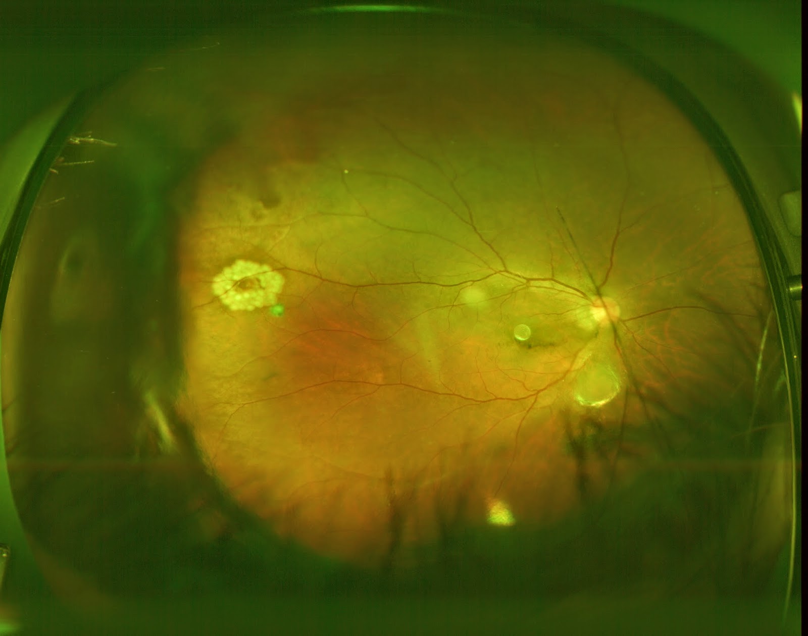
Normal retina is like a thin plastic sheet. This is spread over the inner surface of eye ball like a sticker. Eye ball is filled with vitreous gel which helps in attaching the retina to the surface. In myopia the eye ball is big. The inner surface area is more than normal. To cover this extra inner surface , the retina gets stretched. So the retina in myopes is much thinner and stretched. As the time goes, this thin retina can become friable in certain areas which are called as Retinal degenerations. Later they may tear up causing “Retinal Holes” or “Retinal Breaks”.  If untreated at this stage , these holes can become access points to water in vitreous gel. Once the water enters these breaks, it reaches beneath the attached sticker (Subretinal Space). From here it spreads beneath the retina and detaches it from its surface. This is called as Retinal Detachment (RD). The detached retina loses its food supply and starts dying within hours. Patient loses vision and may become blind for the rest of his life. RD is a serious blinding eye disorder. RD can lead to permanent blindness. This condition can be prevented by timely treatment of retinal breaks.


MYOPIA AND RETINAL DETACHMENT-
FEW QUESTIONS -THEIR ANSWERS:
1.     What are retinal degenerations and retinal breaks?  Why are myopes at  more risk of developing them?
        In myopia, the retina is stretched out and thus is thinner than normal. Such thin retina becomes eroded in certain areas . These weakened areas are called retinal degenerations. These may later break causing a hole in the retina. These are called as retinal breaks or holes.
        Consider a thin cloth. You stretch it with both hands. As you stretch it further, it becomes thinner in certain areas and later gets torn. Retina in Myopes behaves in similar way.
2.     How do I know if I have retinal breaks?
        There is no way that you will know of their presence. There are no symptoms nor indications. Only a retinal surgeon can detect these holes. Occasionally few patients can have floaters or flashes of light when they develop breaks. You should not rely on these symptoms as they are seen in only small percentage of retinal holes.
3.     Can the retinal holes be prevented?
        Absolutely no. The thin retinal nature comes by birth. It cannot be modified. So the holes cannot be prevented. Can u make a dwarf become tall? No. Certain things cannot be prevented or changed. Retinal breaks are one of those.
4.     What are the complications of retinal breaks?
        Retinal breaks if not treated early, can lead to Retinal Detachment and blindness.
5. What is Retinal Detachment?
        Normally the retina is attached like a sticker to the underlying layer. The separation of retina from the inner surface of eye is called as RD. RD is a serious blinding disorder.
6.     Can Retinal Detachment be prevented?
        Certainly yes. It can be prevented by regular retinal screening examination and timely laser barrage to retinal holes.
7.     What is retinal screening and prevention?
        Patients with retinal holes do not have any eye problem or symptoms. It is the  retinal surgeon, who after examination, detects these holes. The examination which he does to detect these holes is called as Retinal Screening. In case of a hole being detected, he does a laser treatment to seal off the holes . This treatment done to prevent RD is called “Prophylaxis” (Preventive treatment ).
        Retinal Holes are like bombs being hidden in your eyes. All myopes are at risk to have these bombs. If not disconnected and removed at right time, the bomb may explode and cause serious damage. Similarly retinal hole if not treated , can explode in the form of RD and cause serious blindness. The test done by Retinal surgeon to detect these bombs is called as Retinal Screening. The laser treatment which he does to disconnect these bombs is called as “Prophylactic LASER barrage” .
8.     Who should undergo retinal screening?
        Any myope with more than -3 Diopter glass power should undergo yearly retinal screening.
9.     How often should retinal screening be done?
        Usually you should get it done every year, Unless your retinal surgeon advices otherwise.
10.   I am a myope with -5 D glass power. I don’t have any problem with my eyes. I have absolutely no symptoms. Should I still undergo retinal examination?
        Absolutely yes. By the time you develop symptoms, you may already have developed RD. Then you may become blind for the rest of your life.
11.    What  is Pupil dilatation?
        Retina is inside the eye ball. Its like a movie screen inside the theater. The only way to look at it is a small window in our eye, which is called pupil. It is like peeping through the window to watch the movie. The more the window is open , the more the view of screen. Normally the pupil is small, allowing us only partial view of retina. Pupil should be widened to give the full view of retina. The process by which pupil is dilated is called as pharmacodilatation. This is done by instilling eye drops. It takes 30 min. to one hour for full dilatation. After the dilatation, persons near vision will get blurred for few hours. Also he has Photophobia (Inability to see light) for few hours. All these are temporary.
12.   What is “Prophylactic Laser Barrage”?It s a treatment done for the retinal Breaks or degenerations, to prevent  Retinal Detachment. It aims at creating a Barrage (Fence/Dam) around the dangerous lesions (The danger areas). LASER is passed onto the retina and it is welded around the lesions. This adhesive fence which is created , blocks the water to spread beneath the retina. Thus it prevents RD.           

It is a minor procedure done in doctor’s room. It is done under topical anesthesia (eye drops). It needs less than 10 minutes. No injection or rest is required. There is no need to stay in hospital. The patient can resume his/ her activities in a couple of hours. It s entirely safe procedure. Usually a single sitting is sufficient. Few people with extensive lesions may require more than one sitting.


                13.   Does laser barrage give life long protection? Is there a need for yearly screening after laser barrage?
        Laser barrage does not prevent formation of holes in other areas. Consider a torn cloth. You stitch it. Does this prevent it in getting torn in other sites? Similarly, laser barrage treats only the existing lesions. Patient still has chance of  developing lesions in other sites. Yearly screening and repeat laser, if required, are very much essential.
14.   I underwent LASIK. Should I still undergo retinal screening?
        Yes. LASIK only removes your glasses. It has no effect on your retinal status. Your thin retina still remains risky. LASIK does not reduce the chance of retinal breaks or RD.
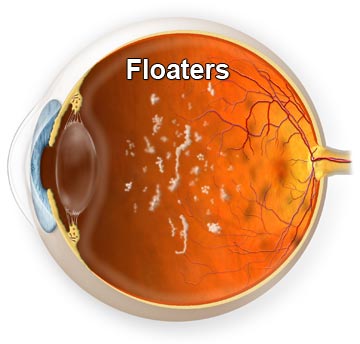
15.   I am a myope. I sometimes see few black spots and strings infront  of my eyes, especially when i look at sky. What are these?
        These are called as “Floaters”. Myopia is a complex disorder with various problems. Glasses is just one of them. Myope have retinal problems. They may also have Vitreous Gel problems. Our eye ball is filled with a Vitreous gel. This gel is usually clear. In some myopes and even in normal people, this gel may get condensed in certain areas , forming opacities. These condensed vitreous strands are seen as spots or strings. These are innocuous and dangerless. But in few cases these floaters may be associated with retinal breaks. So you should undergo retinal screening to detect holes. If no holes are found, then you have nothing to worry. Sometimes you may see Flashes of light, which is called as photopsia. This is again an indication for immediate retinal screening, as it can be associated with retinal breaks.
16.   What are the risk factors for Retinal detachment?
        Myopia is the major risk factor.
        Other risk factors are:
        a.    Previous cataract surgery.
        b.    Severe Injury.
        c.    Previous RD in other eye.
        d.    Family history of RD.
        e.    Retinal degenerations (which can be detected only by a retina surgeon) etc.    
17.   What is the treatment of Retinal Detachment?
        There is no guaranteed treatment for RD. But there are treatments (Complex Vitreoretinal surgeries)  which are aimed at reattaching the retina. But these should be considered as “Damage control measures” rather than guaranteed surgeries. Its like damage control done after a bomb explosion. Our aim should be to prevent the bomb explosion rather than doing damage control after explosion.
        The available treatment modalities for RD are Scleral Buckling and Vitrectomy.
        The type of surgery, type of anesthesia etc are decided by the characteristics of Retinal Detachment.
Scleral Buckle: A flexible band (Scleral buckle) is placed around the eye to counteract the force pulling the retina out of place.The fluid under the retina is drained off if necessary. This is an extraocularoperation with relatively less complications than vitrectomy.

 
Vitrectomy : This is intraocular procedure, where the surgical instruments enter the eyeball. The vitreous is removed, the subretinal fluid is removed and vitreous is replaced with air , fluid or silicon oil.


18.   What are the risks of retinal surgery?
        No surgery is riskless. RD surgery has some risks like Bleeding, Infection, Raised Intraocular pressure, Cataract etc..
        The retina cannot be reattached even with all efforts in some cases
        Most RD surgeries are successful, although a second operation is sometimes needed.
19.   What are the visual expectations after surgery?
        Vision may take many months to improve and in some cases may never return fully. The amount of visual recovery cannot be predicted before surgery. Its only a matter of luck and chance. Unfortunately some patients do not recover any vision.
20.   What happens to the eye if RD surgery is not done?
            It will slowly lose all  existing  vision and becomes totally blind. The eye will eventually become small and shrunken causing an ugly cosmetic appearance.
Retinal Detachment blindness has no cure.
Prevention is the only way.

 
         Regular retinal screening and Timely LASER Barrage is the only way to prevent Blindness.

Thursday, February 23, 2017

Eye flashes after retinal surgery


Eye flashes after retinal surgery could be the craze regarding present day well-known articles, small children with the study of your seo so that you can supply full substance all of us attempt to find pictures connected to all the Eye flashes after retinal surgery . and then the good results you will discover following ought to be examples of the illustrations or photos is a model.

one photo Eye flashes after retinal surgery


Laser Surgery: Retina Eye Before And After Laser Surgery Pictures All things eye Por Yong Ming: Floaters, flashes of light and retina Ahuja Eye Centre - Eye Hospital - Eye Care Centre Retinal DetachmentInterActive Health Eye Floaters Treatment Retinal Specialists of Huntington Beach, CA Displaying 18> Images For - Retinal Detachment Symptoms

Eye flashes after retinal surgery - it has recently been published with the hope of which it is possible to motivate good to a single. The next few paragraphs might offer for a a blueprint if you find yourself bewildered to find the perfect direct The Eye flashes after retinal surgery articles or blog posts may very well be hard possibility to remain utilized on their work schedule, considering that it comes with its own arrange should look and feel a great deal more completely satisfied Eye flashes after retinal surgery - Very beneficial in your case therefore we are trying to find a reliable form which inturn will help uou acquire determination while not frustration. never forget to help you store this site, as its possible in the future you have to pick the software lower back while your current inspirational concepts.